Research

We study the systems developmental biology, biophysics and biomechanics of early spinal column development in zebrafish. More specifically, we seek to understand how interactions among molecules give rise to cell behavior, then how cell behavior gives rise to tissue properties and then how tissue properties give rise to the development of the embryo. Our experimental approach is driven by the idea that quantitative in vivo analysis will lead to fundamental insights into the emergence of biological organization from the collective interaction of its constituent parts. We combine in vivo biophysics, embryology, genetics, live imaging and systems level data analysis and modeling to study pattern formation and morphogenesis. We are an interdisciplinary group with developmental biologists, a marine biologist, a biophysicist and a biomedical engineer.                                                                                                                                             
One area of research focuses on cell migration in the tailbud during body elongation.  We perform live imaging of the tailbud in which all cell nuclei are fluorescently labeled, track the movement of each cell and systematically analyze cell motion.  In this Figure, the cell tracks are segmented into four domains within the tailbud. The general flow of cells is indicated by the yellow arrows in the figure above, and the characteristics of cell motion in each colored domain is briefly described.


We recently described the phenomena of mechanical information in the tailbud as illustrated in the second Figure.  By perturbing cell motion in the tail organizer, we detected long-range alterations in cell motion in the posterior neural tube.  The mechanical information appears to travel too fast to be mediated by gene transcription and translation.  The propagation of mechanical information was modeled in a computer simulation of the tailbud.  In the third figure, an acute perturbation is modeled as a sudden increase in cell repulsion (green) in some cells in the tail organizer.  This simulation depicts a side view of the tailbud. The perturbation leads to a burst of cells in the posterior neural tube that transiently reverse the direction of motion (red).  The mechanical information essentially travels like a traffic jam propagates upstream of an accident.

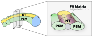
Another area of research in the lab focuses on the role of the extracellular matrix (ECM) in early spinal column development.  As illustrated in the next figure, we found that the ECM protein Fibronectin acts as a “smart adhesive” (red) that binds the posterior neural tube (purple) to the left and right columns of presomitic mesoderm (cyan). The interfaces between these tissues behave as adhesive lap joints, which are commonly used in engineering and woodworking.  Adhesive lap joints are formed by adhering two objects with a glue, and the overlapping domain exhibits a stress profile with stress highest at the outer edges of the overlapping domain.  Fibronectin acts as a smart adhesive in that it constantly remodels as the embryo develops to the region of highest stress. This creates a medial to lateral gradient of Fibronectin fibers between the tissues.  

We imaged Fibronectin matrix remodeling using a photoconvertible Fibronectin transgene.  As illustrated in the next figure, we photoconverted spots of Fibronectin matrix from green to red at the interface between the neural tube and PSM, and followed the deformation of the spots over time.  We found that Fibronectin mediated adhesion resists convergence of the neural tube and predisposes the embryo to spina bifida.  In fact, the neural tube closure defect of the n-cadherin mutant can be rescued by reducing adhesion to the Fibronectin matrix.  However, loss of this cell-Fibronectin adhesion leads to a decrease in bilaterally symmetric morphogenesis of the developing trunk.

Integrins are heterodimers that form an important class of ECM receptors.  The assembly of a Fibronectin matrix is controlled by regulating Integrin activation.  The surface of the PSM is coated in Fibronectin matrix but there is little Fibronectin matrix within the tissue. However, all cells in the PSM express both the Integrin and Fibronectin, so the question arises, why doesn’t the Fibronectin matrix form throughout the tissue?  We found that Fibronectin matrix assembly is inhibited by physical interaction between Integrin heterodimers expressed on adjacent cells within the tissue.  This physical complex includes N-Cadherin which helps stabilize the association between Integrins on adjacent cells.  We measured these interactions in live embryos using Fluorescent Cross-Correlation Spectroscopy (FCCS).  This was the first identification of interactions between Integrins on adjacent cells and was the first measurement of the Kd between Cadherins in adjacent cells.  This regulatory mechanism restricts Fibronectin matrix assembly to the surface of the tissue where the Integrins a free to activate.  More recently, we have adapted a FRET-FLIM assay in the living embryo to measure the conformational change that accompanies Integrin activation.

If you want to listen to what we do see http://www.benefunder.com/causes/402/scott-holley